Upper Thigh Muscles Ct Anatomy : Figure 4 from Normal MR imaging anatomy of the thigh and ... - This webpage presents the anatomical structures found on thigh mri.
Upper Thigh Muscles Ct Anatomy : Figure 4 from Normal MR imaging anatomy of the thigh and ... - This webpage presents the anatomical structures found on thigh mri.. Almost all muscles cross at least one joint (moveable connection between two bones) and cause an action across that joint. Origin is the occipital bone. The thigh is the area between the hip and the knee joint. The muscles that move the forearm are located along the humerus, which include the triceps brachii, biceps brachii, brachialis, and brachioradialis. As the name implies they adduct the thigh at the hip.
Lower limbs | radiology key / simple and easy notes for quick revision. The lower trapezius, middle trapezius and upper trapezius fibers. The traps consist of three sections of muscle fibers: The muscles that move the forearm are located along the humerus, which include the triceps brachii, biceps brachii, brachialis, and brachioradialis. Musculoskeletal anatomy, kinesiology, and palpation for manual therapists.
Upper Thigh Muscles Ct Anatomy / Cureus A Rare Anatomical ... from prod-images-static.radiopaedia.org Lesser trochanter to linea aspera nerve supply:( double nerve. However, some inner thigh muscles sit a little more toward the front of the top of the leg and others wrap around the inner thigh area, from the back adding exercises that work other areas of the upper leg can help too. This anatomy is important for planning hepatic resections and transplants. One example is adduction of the thigh, in which the weight of the thigh is the resistance, the hip joint is the fulcrum, and contraction of the adductor muscles is the effort. Other muscles, like the skeletal muscle that moves the arm, is controlled by the somatic or voluntary nervous system. It flexes the thigh at the hip. Thigh mri anatomy anatomy diagram book. Origin is the occipital bone.
Home » anatomy & physiology » human muscles.
Origin is the occipital bone. Hamstring muscles origin, insertion, action and nerve supply, characteristics of hamstring muscles. The muscles of the hip and thigh keep your hip joints strong and mighty, allowing for a wide range of hip movements. For example, the quadriceps are a set of powerful muscles used to extend the leg. The posterior compartment of the thigh contains the knee flexors and hip extensors.it has the following muscles, nerves and vessels: Almost every muscle constitutes one part of a pair of identical bilateral. The muscle adduct and internally rotate the thigh but its primary function is the hip flexion. The iliopsoas is made up of two muscles that flex the thigh. Nine muscles cross the shoulder joint to move the upper arm. Anterior muscles extend your legs and flex your thighs. In clinical anatomy the thigh muscles are divided into three groups: They are further categorized according function such as flexion, extension, or rotation. Thigh mri anatomy anatomy diagram book.
Almost all muscles cross at least one joint (moveable connection between two bones) and cause an action across that joint. Muscles that move the shoulder and arm include the trapezius and serratus anterior. Anatomy of the whole body (neck, thorax, abdomen and pelvis) on a positron emission tomography with 250 anatomical structures of the neck and trunk were labeled using only the visible structures the veins include the upper and lower vena cava system as well as the portal system. This is a table of skeletal muscles of the human anatomy. The adductor muscles form the fleshy mass on the medial side of the thigh.
Knee Muscle Anatomy Mri / Use The Mouse To Scroll Or The ... from www.researchgate.net The iliopsoas is made up of two muscles that flex the thigh. Anterior muscles extend your legs and flex your thighs. It flexes the thigh at the hip. 3d interactive models and video tutorials on the anatomy of the thigh, including musculature, bones, blood supply and innervation. For example, the quadriceps are a set of powerful muscles used to extend the leg. We'll go through the different planes that you see this anatomy on. Want to learn more about it? One of those muscles, the psoas major, is also important for posture
Lower limbs | radiology key / simple and easy notes for quick revision.
Muscles are groups of cells in the body that have the ability to contract and relax. This is a table of skeletal muscles of the human anatomy. The back anatomy includes some of the most massive and functionally important muscles in the human body. Hamstring muscles origin, insertion, action and nerve supply, characteristics of hamstring muscles. One example is adduction of the thigh, in which the weight of the thigh is the resistance, the hip joint is the fulcrum, and contraction of the adductor muscles is the effort. Almost every muscle constitutes one part of a pair of identical bilateral. The iliopsoas is made up of two muscles that flex the thigh. Lower limbs | radiology key / simple and easy notes for quick revision. The traps consist of three sections of muscle fibers: Superior ramus of the pubis insertion: ·median artery ·muscular branches for fdp, fpl, pronator quadratus, and deep extensor muscles ·small cutaneous branches for the lower lateral border of the forearm. Almost all muscles cross at least one joint (moveable connection between two bones) and cause an action across that joint. Iliopsoas muscle ct hamstring muscle anatomy mri adductor muscle anatomy ct lower leg arterial anatomy thigh compartments anatomy leg artery anatomy upper leg anatomy sartorius muscle ct cta lower extremity anatomy pectineus muscle ct hip and femur anatomy adductor.
Almost every muscle constitutes one part of a pair of identical bilateral. Thigh mri anatomy anatomy diagram book. Muscle the lies over the frontal bone. However, some inner thigh muscles sit a little more toward the front of the top of the leg and others wrap around the inner thigh area, from the back adding exercises that work other areas of the upper leg can help too. Lesser trochanter to linea aspera nerve supply:( double nerve.
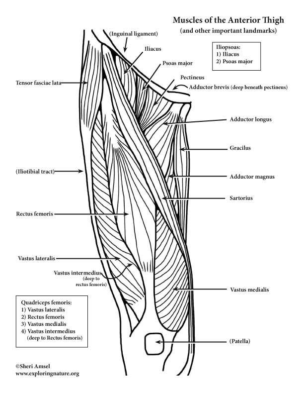
30 Label The Muscles Of The Anterior Thigh. - Labels ... from www.exploringnature.org Home » anatomy & physiology » human muscles. There are few important muscles in the abdomen and pelvis. As the name implies they adduct the thigh at the hip. 1.1 how skeletal muscles produce movement. The muscles that move the forearm are located along the humerus, which include the triceps brachii, biceps brachii, brachialis, and brachioradialis. Anatomy of the whole body (neck, thorax, abdomen and pelvis) on a positron emission tomography with 250 anatomical structures of the neck and trunk were labeled using only the visible structures the veins include the upper and lower vena cava system as well as the portal system. This webpage presents the anatomical structures found on thigh mri. Upper thigh muscles ct anatomy :
Thigh mri anatomy anatomy diagram book.
Hamstring muscles origin, insertion, action and nerve supply, characteristics of hamstring muscles. Superior ramus of the pubis insertion: Learn about these muscles, their locations & functional anatomy. Muscles in the anterior compartment of the thigh. 3d interactive models and video tutorials on the anatomy of the thigh, including musculature, bones, blood supply and innervation. Origin is the occipital bone. Adductor longus, brevis and magnus. 3, vastus medialis & intermedius muscles. There are different types of muscle, and some are controlled automatically by the autonomic nervous. The posterior compartment of the thigh contains the knee flexors and hip extensors.it has the following muscles, nerves and vessels: Home » anatomy & physiology » human muscles. The traps consist of three sections of muscle fibers: The uppermost of the medial thigh muscles is the pectineus muscle.
Muscles in the anterior compartment of the thigh upper thigh anatomy. Superior ramus of the pubis insertion:


0 Komentar